We’ve rebranded some of our products, learn more ›
CODEX® is now PhenoCycler, Phenoptics™ is now Phenolmager.
More Signatures, Faster than Ever!
Spatial Signatures are a new class of highly predictive biomarkers that more accurately predicted patient response to immunotherapies than current biomarker assays.
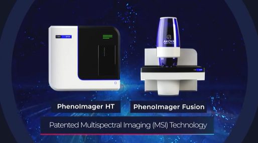
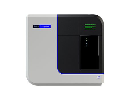
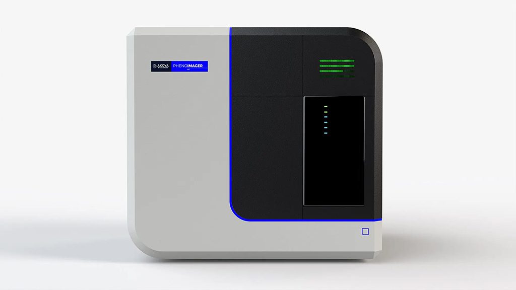
PhenoImager® HT 2.0 is the fastest solution for the development of spatial signatures, powered by Akoya’s patented Multispectral Imaging (MSI) and spectral unmixing technologies that enables rapid and accurate spatial phenotyping at scale.
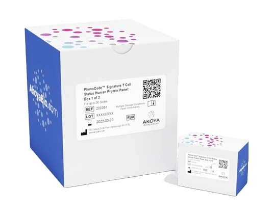
Pave your path to spatial signatures with PhenoImager HT 2.0 and the newly launched PhenoCodeâ„¢ Signature Panels.
Why choose PhenoImager HT 2.0?
Speed
The fastest imager for spatial phenotyping and signature development, HT 2.0 supports walk-away automation with a straightforward and user-friendly workflow enabling you to scale projects effortlessly.
Accuracy
A new onboard spectral unmixing process that runs seamlessly in parallel with imaging means never having to compromise on data quality, regardless of your project size.
Throughput
HT 2.0 is ideally suited for an automated mIF staining and imaging workflow, providing the capacity to image over 400+ multiplex stained slides per week to fit any project of your scale.
Proven
Akoya’s technologies have been cited more than a 1,000 times. With the installation of our 1000th instrument, we are proud to be one of the most trusted partner in the field of spatial biology.
Spatial Signatures
The Next Generation of Predictive Biomarkers
What are Spatial Signatures?
Spatial Signature are predictive biomarkers based on spatial relationships and protein co-expression of specific cellular subsets assessed within the context of the tumor micro-environment (TME). These spatially determined predictive biomarkers provide deeper insights into the tumor-immune biology and could inform treatment response.
Spatial Signatures can be based on the following within the TME (tumor, stroma, invasive margin): presence or absence of a phenotype, proximity, density, structures or unique neighborhood or a combination of these features.
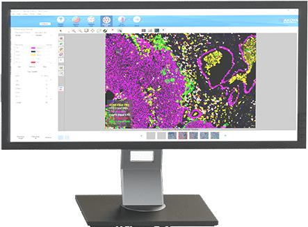
Visualize
Quantify
Predict

How are Spatial Signatures measured?
At the core of spatial signature development is multiplexed immunofluorescence (mIF), a next-generation pathology workflow that generates quantitative and reproducible insights about tumor samples.
What is the best platform for the development of Spatial Signatures?
The development of accurate and reproducible spatial signatures requires a reproducible and robust solution.
The MITRE study, is the first multi-institutional analytical demonstration of a spatial biology workflow and establishes the reproducibility and robustness of Akoya Biosciences’ PhenoImager platform for spatial signature development.
Case Study: A framework for developing a robust Spatial Signature
In this study, the authors present a detailed framework for mIF assay development and data analysis on the PhenoImager platform that can be used to standardize results across research institutions – an important step towards generating highly predictive spatial signature for clinical implementation.
Application: Leveraging Spatial Signatures for Companion Diagnostics
A first-of-its-kind companion diagnostic, the OncoSignature® test, developed by Acrivon, will leverage the spatial phenotyping capabilities of the PhenoImager solution to localize and quantify the expression of a signature of clinically relevant protein biomarkers within the tumor during clinical development.
Explore Data
Hear from Our Users
PhenoImager Technology
Watch this video to learn about how the PhenoImager instruments use a patented Multispectral Imaging (MSI) and spectral unmixing technology to isolate autofluorescence and biomarkers of interest regardless of their intensity or any spectral overlap into separate channels, providing accurate and quantitative data for spatial phenotyping and spatial signature development.
A Simple End-to-End Workflow
Stain
Image​​
Analyze​​